Male reproductive system has two parts viz. The reproductive tract and accessory glands. The main reproductive organ or male reproductive organ or male gonad is testis which is situated in scortum.
Main reproductive tract :
1. Scrotum : The scrotum is a pouch of pigemented skin arising from lower abdominal wall and having wall consisting of dartos tunic which are smooth muscles . It is divided into two compartments right and left by muscle septum . Scrotum protects testes and acts as thermoregulator , i.e., regualtes the temperature for proper funtioning of testis
2 . Tests :
(a) Tests are primary sex that are soft smooth , pinkish oval having dimensions of about 4.5 cm length , 2.5 cm width and 3 cm thickness.
(b) Tests ate extra abdominal in position . They are suspended in the scrotum by the spermatic cord and is connected to the wail of scrotum by short fibromuscular band called gubernaculum.
(c) There are about 200 to 300 lobules in each testis in which there are 1 to 4 convoluted loops composed of germinal epithelial cells . These are called seminiferous tubules.
(d) Seminiferous tubules converag towards posterior surface and form a network of irregualr tubules called rete testis .
(e) Seminiferous tubules converge of spermatogonia ( sperm mother cells ) and nurse cells or cells of Sertoil .
(f) Between seminiferous tubules there are inteestitial cells or cells of Leydig which secrete the hormone testosterone after puberty .
3. Vasa efferntia : Vasa efferentia are a pair of ducts starting from the rete testis and entering in epididymis . They are 15 to 20 fine convoluted ductules that pierce the tunica albuginea to enter the caput epididymis .
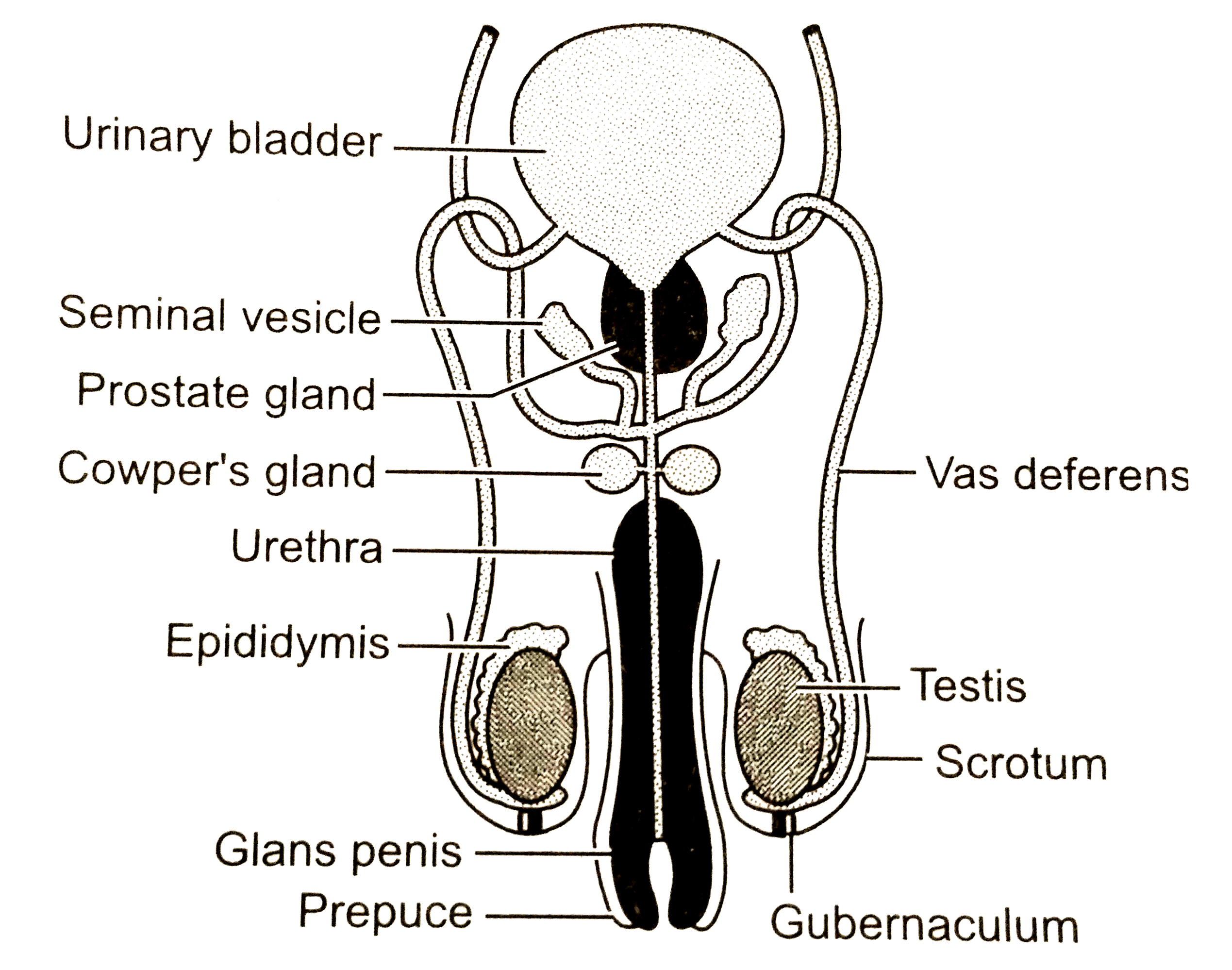
4. Epididymis : Epididymis are 'C' shaped paired structure showing about 6 metres long highly coiled duct situated on the posterior border of each testie . It is differentiated into the following three regions :
(a) Caput epididymis which is upper wider head that receives vasa efferentia . Here the sperms undergo physiological maturation acquiring increased motility and fertilizing capacity .
(b) Corpus epididymis which is middle narrower body .
(b) Corpus epididymis which is middle narrower body .
(c) Cauda epididymis which is a lower duct or tail , sperms remain for short period and then enter the vas deferens.
5. Vasa defernia : (a) A pair of tubular structures about 40 cm long arising from cauda epididmis are called vasa deferentia.
(b) Each vas deferens enters the abdominal cavity through the inguinal canal and then ascends in the form of spermatic cord, medially towards the posterior wall of the urinary bladder .
(c) Vas deferens of each side is joined by the duct from seminal vesicle to form ejaculatory duct .
6. Ejaculatory duct : About 2 cm long pair of ducts formed by joining of vas deferns and a duct of seminal vesicle are the ejaculatory ducts. Both ejaculatory ducts open into urethra near the prostate glad . Seminal fluid containing spermatozoa are carried by ejaculatory duct to the urethra.
7. Urethra : The male urethea is a common pathway for the flow of urine and semen. The urethra has three parts , viz ., prostatic urethra , membranous urethra and penile uretha. Urethra carries both urine and semen . It has two sphincters - internal sphincter made up of smooth muscle fibres at its beginning and external sphincter made up of striated muscles.
8. Penis :
(a) Penis is the copulatory organ used for insemination or deposition of sperms in female genital tract.
(b) It is cylindrical ,erectile and pendulous organ suspended in public region in front of scrotum . Through the length of penis passes the uretha .
(c) It contains three column of erectile tissues which has abundant blood sinuses.
(d) The penis contains two postero - lateral tissue called corpora cavernosa and a median corpus spongiosum .
(e) The urethra passes through corpus spongiosum . Hence , it is also called spongia urethra .
(f) Near the tip of the penis , the corpus spongiosum is enlarged to form a soft and highly sensitive glans penis. It is covered by a loose retractable fold of skin called prepuce of foreskin .
Accessory sex glands :
1. Seminal vesicles :
(a) The semina, vesicals are two small fibromuscular pouches present on the posterior side of the urinary bladder .
(b) They have semina ducts which join with vas deferens and form ejaculatory ducts.
(c) Seminal fluid is secreted by seminal vesicle which forms about 60% of the total volume of the semen .
(d) Seminal fluid is a viscous fluid containing fructose , fibrinogen and prostaglandins . Fructose provides energy to sperms for swimming .
(e) The prostaglandins stimulate contractions in the female reproductive tract to help the process of fertilisation .
(f) The fibrinogen helps in coagulation of semen after ejaculation .
2. Prostate gland :
(a) About 20 to 30 seprate lobes form a prostate gland which opens separately into the urethra.
(b) It secretes prostatic fluid which is about 30% of total volume of semen .
(c) The prostatic fluid is a whitish and alkalin liquid .
(d) The prostatic secretion neutralizes the acidity of vaginal secretion .
(e) Due to its alkalinity sperms become motile and thus it facilitates the process of fertilization .
3. Cowper's glands (Bulbo-urethral glands ):
Situated in either sides of membranous urethra are pea - sized Cowper's glands which secrete an alkaline viscous fluid .


