Home
Class 12
BIOLOGY
The figure shows a diagrammatic represen...

The figure shows a diagrammatic representation of the sectional view of cochlea with labels A, B, C and D. Select the option with correct identification and main characteristic or function:

A

D-Scala tympani-filled with endolymph

B

C -Scala media-filled will! perilymph

C

B -Hair cells-pressing against the tectorial membrane as a result nerve impulses are generated

D

A -Reissnpr's membrane-responsible for causing auditory reflex.

Text Solution

Verified by Experts

The correct Answer is:
C
Promotional Banner

Topper's Solved these Questions

  • SENSORY ORGANS

    MOTION|Exercise EXERCISE - 2|7 Videos
  • RESPIRATION IN PLANTS

    MOTION|Exercise EXERCISE - 4|19 Videos
  • SEXUAL REPRODUCTION IN FLOWERING PLANTS

    MOTION|Exercise Exercise - 4(AIIMS-2018)|25 Videos

Similar Questions

Explore conceptually related problems

the figure shows a diagrammatic view of human respiratory system with labels A, B , C and D . Select the option which given correct identification and main function and / or characteristic

Amongst labels a - d of human urinary system, select option with correct identification and characteristics/function

The figure given here shows diagrammatic representation of internal organs of frog Identify A to E and select the correct option

The figure shows ribs and rib cage labelled with A,B,C and D. select the option which gives correct identification :-

The figure given here shows diagrammatic representation of internal organs of frog. Identify A to E and select the correct option :

The figure below shows development of follicles (A, B, C, D). Select the option giving correct identification together with its function?

Refer to the given diagrammatic representation of an electron micrograph of a section of chloroplast and answer the Select the option that correct identifes X,Y and Z.

Diagrammatic representation of trophic levels in an ecosystem is given below. Identify A, B, C and D by selecting correct option:

MOTION-SENSORY ORGANS-EXERCISE - 3
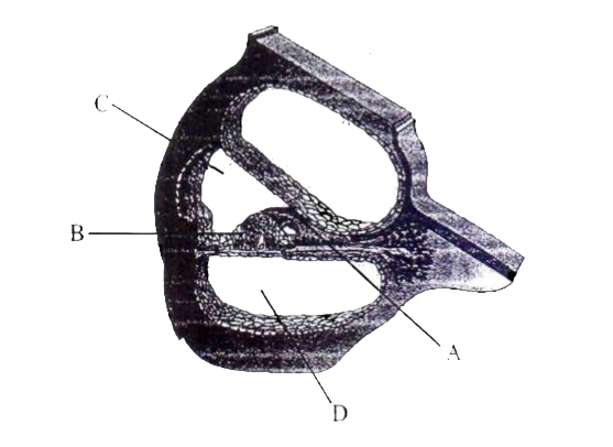
  1. Identify A, B, C, D in the given diagram. Which of the following among...

    Text Solution

    |

  2. For the given diagram which labelling and function is correctly matche...

    Text Solution

    |

  3. The figure shows a diagrammatic representation of the sectional view o...

    Text Solution

    |

  4. Assertion. Tympanic membrane separates the external ear from the middl...

    Text Solution

    |

  5. Assertion :- Cornea causes the maximum refraction of the light rays. T...

    Text Solution

    |

  6. Assertion :- The receptor cells in the organ of Corti transmit impulse...

    Text Solution

    |

  7. Assertion :- Only the pigmented layer of the neuro sensory tunic conti...

    Text Solution

    |

  8. Assertion :- Main function of ear is body balance. Reason:- Organ o...

    Text Solution

    |

  9. Assertion :- Inverted & real image is formed on retina. Reason:- Aq...

    Text Solution

    |

  10. Assertion :- A real and inverted image is obtained on the retina. R...

    Text Solution

    |

  11. Assertion :- Some person unable to see in the dark. Reason:- They l...

    Text Solution

    |

  12. Assertion :- Blind spot of the retina of the eye is devoid of the abil...

    Text Solution

    |

  13. Assertion :- Accomodation is done by Iris of eye. Reason:- Lens is ...

    Text Solution

    |

  14. Assertion :- Accomodation power is present in human eye. Reason:- It...

    Text Solution

    |

  15. Assertion :- If we remove hair cell, then hearing is not possible. R...

    Text Solution

    |

  16. Assertion :- Vision of eye is not disturbed in "Red eye" . Reason:- ...

    Text Solution

    |

  17. Read the assertion and reson carefully to mark the correct option out ...

    Text Solution

    |

  18. Assertion :- The auditory ossicles help in hearing. Reason:- Auditor...

    Text Solution

    |

  19. Assertion :- Organ of Corti rests on tectorial membrane having hair ce...

    Text Solution

    |

  20. Assertion :- No image formation occurs at retina where the optic nerve...

    Text Solution

    |