Home
Class 12
BIOLOGY
Draw the diagram of a microsporangium an...

Draw the diagram of a microsporangium and lable its wall layers.Write briefly about the wall layers?

Text Solution

Verified by Experts

The diagram representation of a microsporangium is shown below

In a transverse section a typical microsporangium is circular in outline and is surrounded by four wall layers.
(a) Epidermis : The epidermis is the outermost protective layer. It is composed of tangentially flattened cells . The cells are closely fitted and heve thick walls which is helpful in the dehiscence of anther.
(b) Endothecium it is present below the epidermis and expands radically with febrous thickenings, at maturity these cells loose water, at contract and help in dehiscence of pollen sac.
(c ) Well Layers it is present between well marked endothecium and tapetum. These are thin walled layers, arranged in one to five layers, which also help in dehiscence of anther.
(d) Tapetum it is the innermost wall layer with large cells ,thin cell walls abundent cytoplasm and have more than one nucid . Tapetum is a nutritive tissue which nourishes the developing pollen grains.
The centre of the microsporangium consists of sporogenous tissue which undergoes meiotic divisions to form microspore tetrads. This process is known as microsporogensis.
Promotional Banner

Topper's Solved these Questions

  • REPRODUCTIVE HEALTH

    NCERT EXEMPLAR|Exercise Reproductive Health|37 Videos
  • STRATEGIES FOR ENHANCMENT IN FOOD PRODUCTION

    NCERT EXEMPLAR|Exercise Strategies For Enhancment In Food Production|77 Videos

Similar Questions

Explore conceptually related problems

Draw the diagram of a microsporangium and lebel its wall parts. Write briefly on role of endothecuium.

Middle layers of the microsporangial wall

The innermost wall layer of microsporangium is called.

Inner most jacket layer of microsporangium is:

The first wall layer of cell is

Wall is two -layered in

The innermost wall layer of anther

The nutritive layer of pollen sac/microsporangium is

Innermost microsporangial wall layer that nourishes pollen grains is

Draw and label the enlarged view of microsporangium of an angiosperm . State the function of its innermost wall layer.

NCERT EXEMPLAR-SEXUAL REPRODUCTION IN FIOWERING PIANTS-Sexual Reproduction In Fiowering Piants
  1. Which is the triploid tissue in a fertilised ovule? How is the triploi...

    Text Solution

    |

  2. Are pollination and fertilisation necessary in apomixis? Give reasons.

    Text Solution

    |

  3. Identify the type of carpel with the help of diagrams given below

    Text Solution

    |

  4. How is pollination carried out in water plants?

    Text Solution

    |

  5. What is the function of the two male gametes produced by each pollen g...

    Text Solution

    |

  6. List three strategies that a bisexual chasmogamous flower can evolve t...

    Text Solution

    |

  7. Given below are the events that are observed in an artificial hybridis...

    Text Solution

    |

  8. Vivipary automatically limits the number of offsprings in a litter. Ho...

    Text Solution

    |

  9. Does self-incompatibility impose any restrictions on autogamy? Give re...

    Text Solution

    |

  10. In the given diagram, write the names of parts shown with lines.

    Text Solution

    |

  11. What is polyembryony and how can it be commercially exploited?

    Text Solution

    |

  12. Are parthenocarpy and apomixis different phenomena ? Discuss their ben...

    Text Solution

    |

  13. Why does the zygote begin to divide only after the division of primary...

    Text Solution

    |

  14. The generative cell of a two celled pollen divides in the pollen tube,...

    Text Solution

    |

  15. In the figure given below label the following parts-male gametes, egg ...

    Text Solution

    |

  16. Starting with the zygote, draw the diagrams of the different stages of...

    Text Solution

    |

  17. What are the possible types of pollinators in chasmogamous flowers. Gi...

    Text Solution

    |

  18. With a neat, labelled diagram, describe the parts of a mature angiospe...

    Text Solution

    |

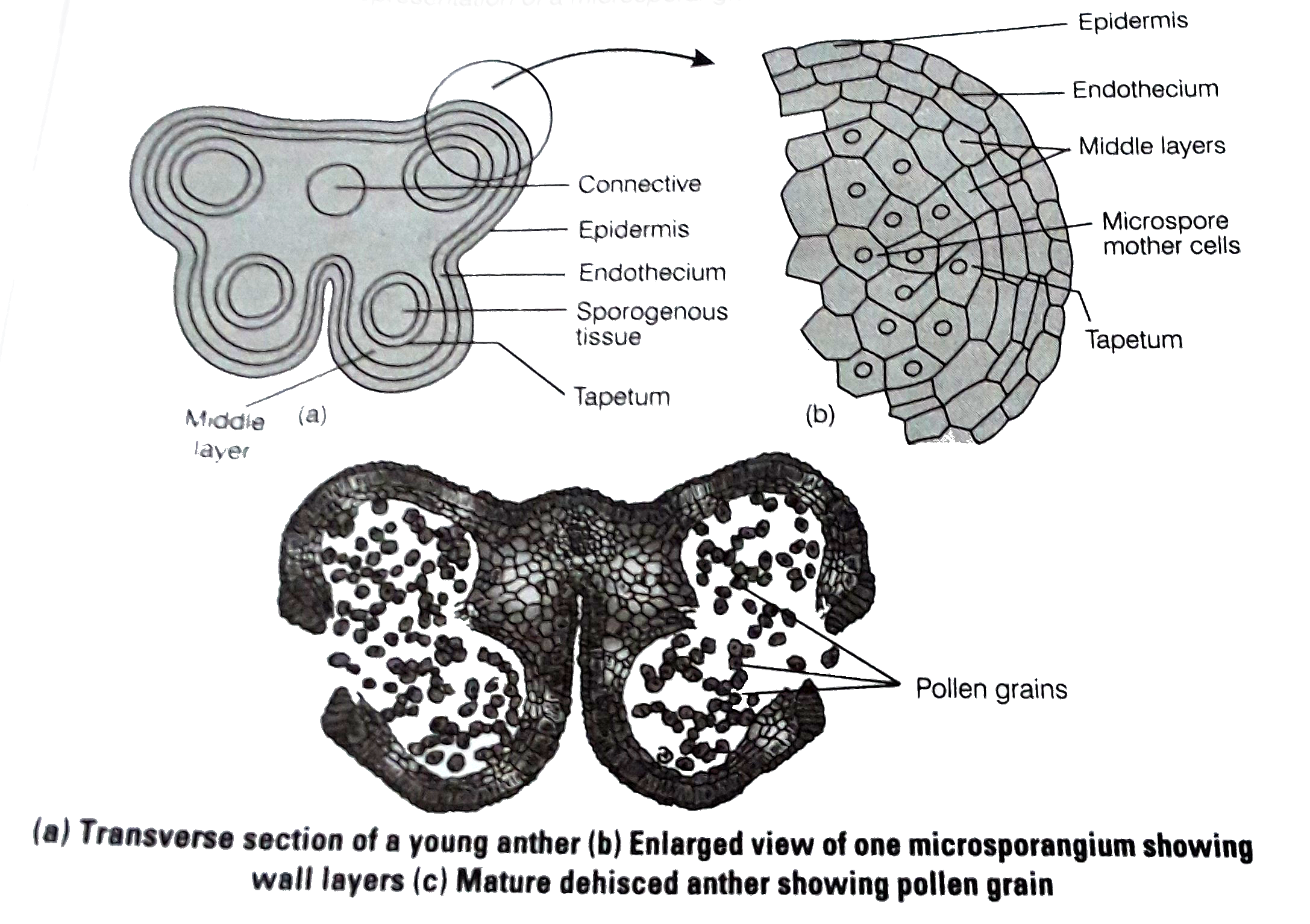
  19. Draw the diagram of a microsporangium and lable its wall layers.Write ...

    Text Solution

    |

  20. Can an unfertilised, apomictic embryo sac give rise to diploid embryo ...

    Text Solution

    |