Home
Class 9
BIOLOGY
(a) Tarun was asked to draw a diagram of...

(a) Tarun was asked to draw a diagram of sclerenchyma as seen in longitudinal section and label it . His figure is given below. Point out the mistake in the figure.

(b) Are the cells of sclerenchyma live or dead ?

Text Solution

Verified by Experts

(a) The figure is transverse section instead of longitudinal section.
(b) The cells of sclerenchyma are dead cells.
Promotional Banner

Topper's Solved these Questions

  • TISSUES

    PRADEEP|Exercise Long Answer Questions|15 Videos
  • TISSUES

    PRADEEP|Exercise Additional Questions (very short answer questions)|46 Videos
  • TISSUES

    PRADEEP|Exercise NCERT Exampler Problems|33 Videos
  • THE FUNDAMENTAL UNIT OF LIFE

    PRADEEP|Exercise ANALYTICAL|11 Videos

Similar Questions

Explore conceptually related problems

Mohit was asked to draw a diagram of sclerenchyma as seen in longitudinal section and label it. His diagram is as shown. Find out the mistake he has made in the diagram.

For the circuit shown in figure given below, the equivalent resistance points A and B is

Identify the labels A , B , C , and D in the figure given below

Study the given diagram and answer the questions given below: Tegmen develops from the part labelled C in the figure is called

The following diagram opposite illustrates a longitudinal section through a shoot apex. Which of the figures gives below shows the correct appearance of this shoot apex at the formation of the next leaf primordium

Study the given figure and identify the cells labelled as A, B, C and D

Examine the figure given below and select the correct option for labelled parts, a,b,c,d

(a) A student observed a nerve cell and drawn the following figure. He did a mistake in labelling. Find out the mistake (b) Where would you find nerve cells ? What is the function of nerve cell ?

The diagram of the ultranstructure of a plant cell is given below. Identify the functions of the organelles labelled. A,B,C,D,E in the diagram

There are two large identical open tanks as shown in figure. In tanks 1 there is a small hole of cross sectional area A at its base. Tank II has a similar hole, to which a pipe of length h has been connected as shown. The internal cross sectional area of the pipe can be considered to be equal to A. Point 1 marked in both figures, is a point just below the opening in the tank and point 2 marked in both figures, is a point h below point 1 (In fig II, point 2 is just outside the opening in the pipe) (a) Find the speed of flow at point 2 in both figures. (b) Find the ratio of speed of flow at point 1 is first the ratio of speed of flow at point 1 is first figure to that in second figure. (c) Find the difference in pressure at point 1 in both figures.

PRADEEP-TISSUES-Short Answer Questions
  1. What is phloem ? Write its components and functions.

    Text Solution

    |

  2. Write brief note on- (i) Adipose tissue (ii) Areolar tissue.

    Text Solution

    |

  3. Describe structure and functions of bone.

    Text Solution

    |

  4. Draw diagram of neuron and briefly write about its components.

    Text Solution

    |

  5. Answer the following : (i) Which skeletal tissue shows unidirection...

    Text Solution

    |

  6. Write these similarities and three dissimilarities between ligaments a...

    Text Solution

    |

  7. Briefly explain the following : (a) Ligament (b) Tendon (c) Neuron.

    Text Solution

    |

  8. Give functions of the following : (i) Blood platelets (ii) Sieve t...

    Text Solution

    |

  9. What is a cartilage ? How is it different from a bone ?

    Text Solution

    |

  10. (a) Which parts of the body are composed of nerve cells? (b) Write ...

    Text Solution

    |

  11. (i) Study the prepared slide of given plant tissue. Give one character...

    Text Solution

    |

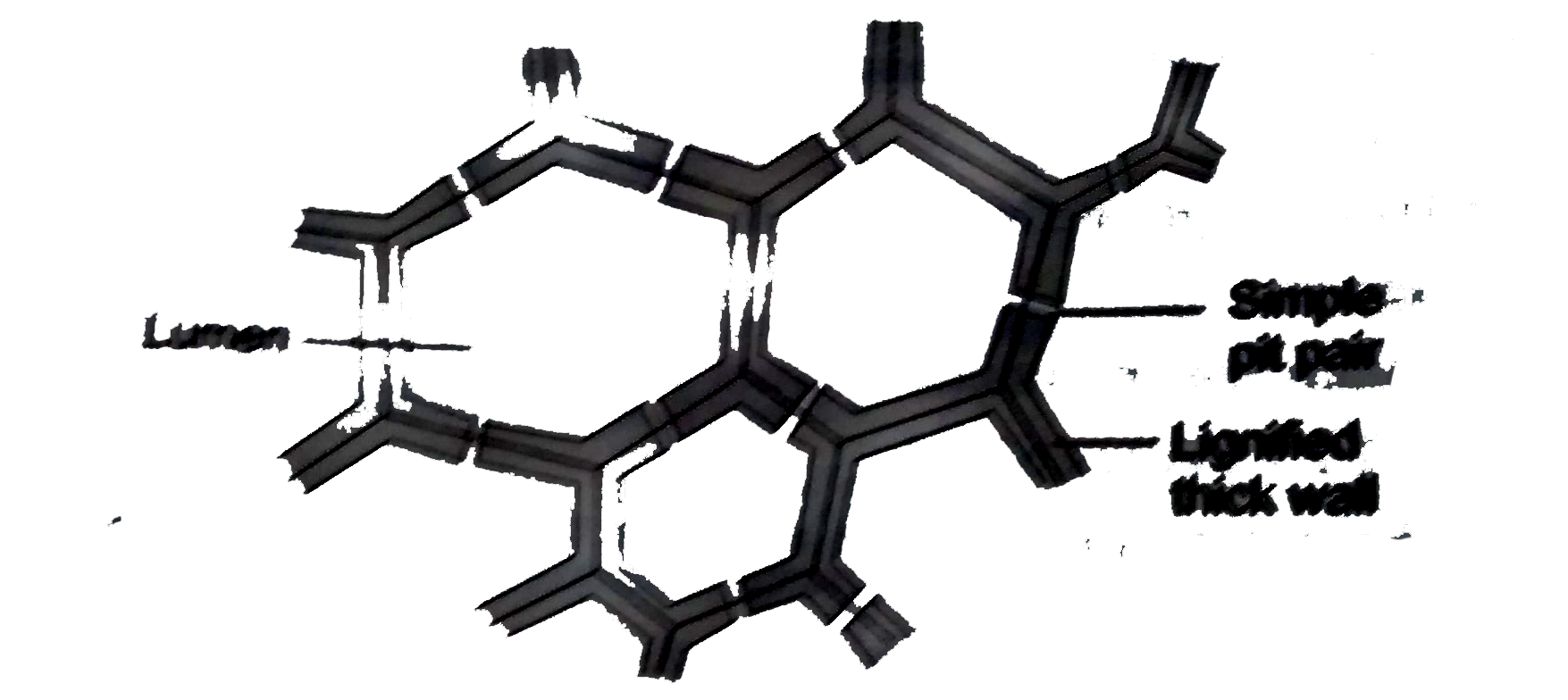
  12. Study the given slide of sclerenchyma plant tissue and answer the foll...

    Text Solution

    |

  13. (a) You have been asked to study the given slide of plant tissue. Give...

    Text Solution

    |

  14. (a) Tarun was asked to draw a diagram of sclerenchyma as seen in longi...

    Text Solution

    |

  15. (a) Two slides were observed under the microscope for spot test as sho...

    Text Solution

    |

  16. (a) A student observed a nerve cell and drawn the following figure. He...

    Text Solution

    |

  17. 1. List three parts of nerve cell. 2. In which part of nerve cell, N...

    Text Solution

    |

  18. (a) Observe slide of smooth muscle fibre under the microscope and draw...

    Text Solution

    |

  19. (a) View prepared slide of cardiac muscle fibres and draw its well lab...

    Text Solution

    |

  20. Study the prepared slide of striated muscles fibres and answer the fol...

    Text Solution

    |