Text Solution
Verified by Experts


Topper's Solved these Questions
BODY FLUIDS AND CIRCULATION
FULL MARKS|Exercise ADDITIONAL QUESTIONS SOLVED ( Fill in the Blanks)|76 VideosBIOMOLECULES
FULL MARKS|Exercise ADDITIONAL QUESTIONS SOLVED (Higher Order Thinking Skills)|16 VideosCELL CYCLE
FULL MARKS|Exercise ADDITIONAL QUESTIONS SOLVED (Higher Order Thinking Skills)|12 Videos
Similar Questions
Explore conceptually related problems
FULL MARKS-BODY FLUIDS AND CIRCULATION-ADDITIONAL QUESTIONS SOLVED ( Answers the following Questions)
- Explain the composition of blood.
Text Solution
|
- Explain the ABO blood groups.
Text Solution
|
- Tabulate the agglutinogens and agguitinins present in the different gr...
Text Solution
|
- Explain Rh factor in brief.
Text Solution
|
- Explain the process of coagulation of blood.
Text Solution
|
- Explain the structure of blood vessels.
Text Solution
|
- Write a short note on coronary blood vessels.
Text Solution
|
- Compare the chambers of heart and the methods of circulation in fishes...
Text Solution
|
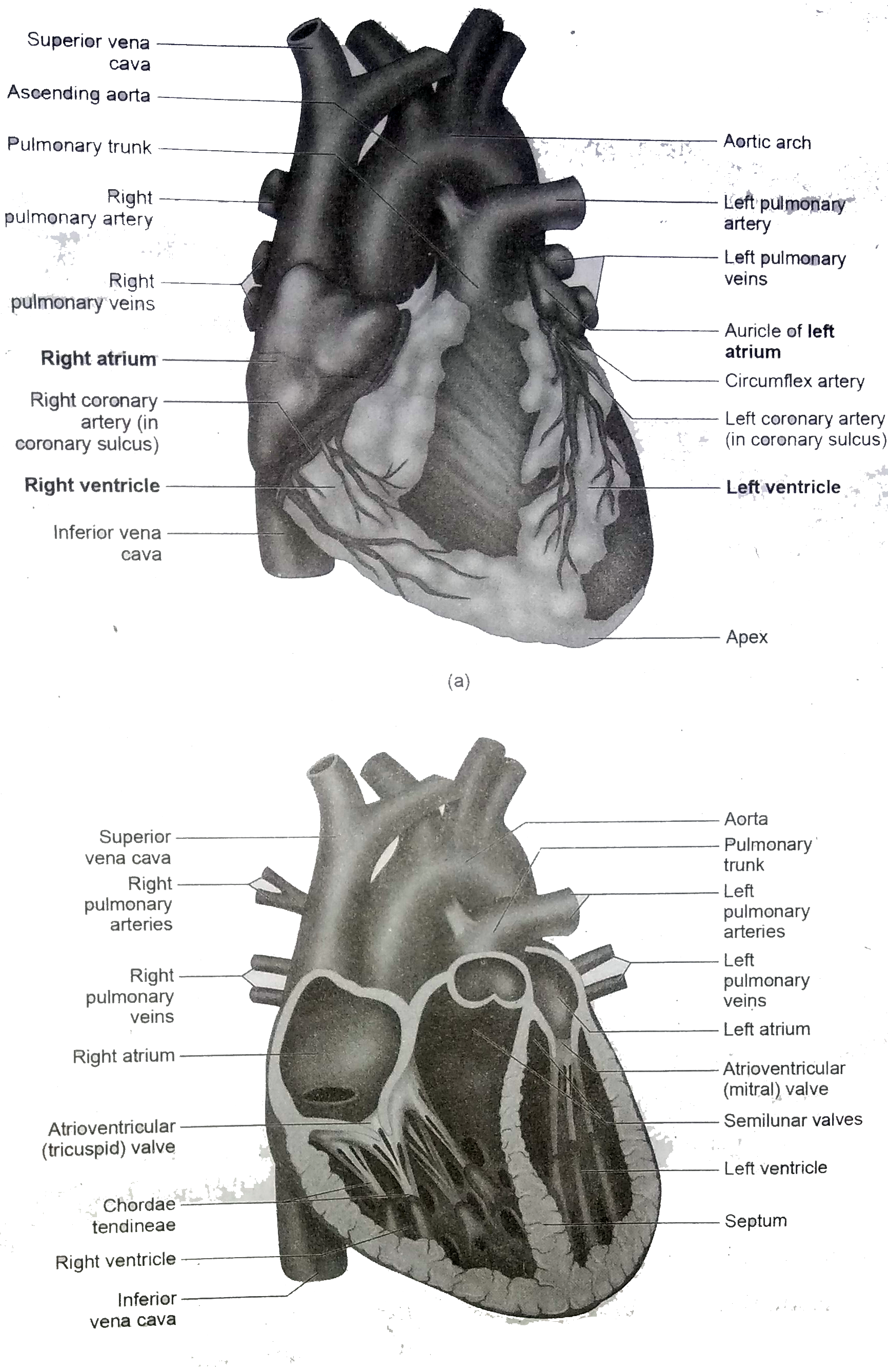
- Draw a labelled diagram of the internal structure of Human Heart.
Text Solution
|
- Explain the cardiac cycle.
Text Solution
|
- What is cardiac output?
Text Solution
|
- Explain the importance of blood pressure.
Text Solution
|
- Explain the recording of electrocardiogram.
Text Solution
|
- Explain the regulation of cardiac activity.
Text Solution
|
- Define:- Hypertension
Text Solution
|
- Coronary heart disease is due to
Text Solution
|
- Define:- Stroke
Text Solution
|
- Define:- Myocardial infarction.
Text Solution
|
- Explain the disorder of rheumatoid heart disease
Text Solution
|
- What is Cardio Pulmonary Resuscitation (CRR) ?
Text Solution
|