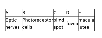
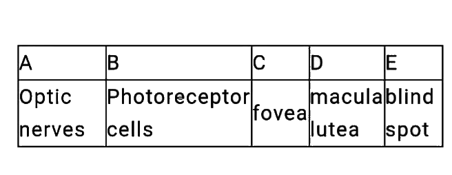
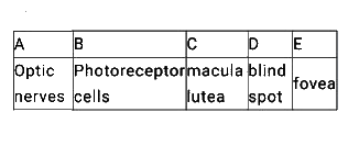
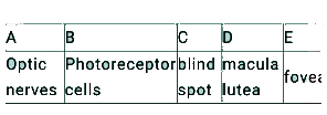
Home
Class 12
BIOLOGY
The A leaves the eye and the retinal blo...

The A leaves the eye and the retinal blood vessels enter it at a point medial to and slightly above the posterior pole of the eyeball. B is not present in that region and hence it is called the C . At the posterior pole of the eye lateral to the C there is a yellowish pigmented spot called D with a central pit called the E

A

B

C

D

Text Solution

AI Generated Solution

The correct Answer is:
To solve the question step by step, we will identify each component mentioned in the question and relate them to the anatomy of the eye. ### Step-by-Step Solution: 1. **Identify A**: The first part of the question states that "A leaves the eye and the retinal blood vessels enter it at a point medial to and slightly above the posterior pole of the eyeball." - **Answer**: A refers to the **optic nerve**. The optic nerve is formed by the axons of the ganglion cells in the retina and exits the eyeball at the optic disc, which is located slightly medial and above the posterior pole. 2. **Identify B**: The next part mentions "B is not present in that region and hence it is called the C." - **Answer**: B refers to **photoreceptor cells** (rods and cones). The optic disc (where the optic nerve exits) does not contain any photoreceptor cells, which is why this area is referred to as the **blind spot** (C). 3. **Identify D**: The question then states that "At the posterior pole of the eye lateral to the C there is a yellowish pigmented spot called D." - **Answer**: D refers to the **macula lutea**. This is a yellowish spot located laterally to the blind spot (optic disc) and is responsible for high acuity vision. 4. **Identify E**: Finally, it mentions "with a central pit called the E." - **Answer**: E refers to the **fovea centralis**. The fovea is a small depression in the macula lutea that contains a high concentration of cone cells, providing the sharpest vision. ### Summary of Answers: - A: **Optic nerve** - B: **Photoreceptor cells** - C: **Blind spot** - D: **Macula lutea** - E: **Fovea centralis** ### Final Answer: The complete answer to the question is: - A = Optic nerve - B = Photoreceptor cells - C = Blind spot - D = Macula lutea - E = Fovea centralis
Promotional Banner

Similar Questions

Explore conceptually related problems

Fovea in the eye is a central pit in the yellowish pigmented spot called

The yellowish pigmented spot at the posterior pole of human eye lateral to lblind spot is

Assertion: The optic nerve leaves the eye at the point called blind spot Reason: Only cones are densely packed at the blind spot, hence is the name.

Read the following statements (A to D) and select the one option that contains both correct statements. A. Z-line is present in the centre of the light band. B. Thin filaments are firmly attached to the M-line. C. The central part of thick filaments, not overlapped by thin filaments is called Z-band. D. Light band contains only thin filaments.

The stress strain graph for a metal wire is shown in the fig. Up to the point E, the wire returns to its original state O along the curve EPO, when it is gradually unloaded. Point B corresponds to the fracture of the wire. (a) Up to what point on the curve is Hooke's law obeyed? (this point some times called proportional limits). (b) Which point on the curve corresponding to elastic limit or yield point of the wire? (c ) Indicate the elastic and plastic regions of the stress-strain graph. (d) Describe what happens when the wire is loaded up to a stress corresponding to the point A on the graph and then unloaded gradually. In particular explain the dotted curve. (e) What is peculiar about the portion of the stress- strain graph from C to B? Up to what stress can the wire be subjected without causing fracture?