A
B
C
D
Text Solution
Verified by Experts


Topper's Solved these Questions
TISSUES
BETOPPERS|Exercise SUMMATIVE WORKSHEET(I)|10 VideosTISSUES
BETOPPERS|Exercise SUMMATIVE WORKSHEET(II) (Fill in the blank)|11 VideosTISSUES
BETOPPERS|Exercise SUMMATIVE WORKSHEET(VIII)|4 VideosRESPIRATION AND EXCRETION
BETOPPERS|Exercise SUMMATIVE WORKSHEET ( ANSWER THE FOLLOWING QUESTIONS)|16 Videos
BETOPPERS-TISSUES-CONCEPTIVE WORKSHEET
- The cells of the tissue that transmit stimulus are known as. The infor...
Text Solution
|
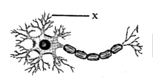
- The given illustration represents a neuron. In the given illustra...
Text Solution
|
- The given illustration represents a neuron. In the given illustra...
Text Solution
|
- The given illustration represents a type of muscle fibre. The ill...
Text Solution
|
- Involuntary muscles control the movement of food in the alimentary can...
Text Solution
|
- The cells of voluntary muscles are long, cylindrical, i and ii. The i...
Text Solution
|
- Which of the following statements about bone is incorrect?
Text Solution
|
- Which of the following statements about blood is incorrect?
Text Solution
|
- The tissue found between the skin and muscles is called i tissue. Fats...
Text Solution
|
- Cartilage has a solid matrix that is composed of i and ii . The info...
Text Solution
|
- Which connective tissue transports gases, nutrients, and wastes to dif...
Text Solution
|
- Ligaments that connect two i are a type of ii tissue. The informatio...
Text Solution
|
- What is the primary function of cuboidal epithelium?
Text Solution
|
- The given figure represents a cell present in the inner lining of the ...
Text Solution
|
- Which illustration represents stratified squamous epithelium?
Text Solution
|
- Which epithelial tissue forms the lining of the mouth?
Text Solution
|
- The linings of i and the ducts of salivary glands are formed by ii epi...
Text Solution
|
- Which type of epithelium is present in the inner lining of the intesti...
Text Solution
|
- Simple squamous epithelial cells are extremely i and ii. The informa...
Text Solution
|
- The xylem is a complex permanent tissue, which is made of four differe...
Text Solution
|