Home
Class 12
BIOLOGY
Describe the structure of mammalian test...

Describe the structure of mammalian testis with the help of labelled diagram.

Text Solution

Verified by Experts

Testes. They are the primary sex organs which develop in the abdominal cavity during early foetal life. They descend into the scrotum through inguinal canals during 7th month of development. There is a pair of testes that are suspended in the scrotum by the spermatic cords. A fibrous cord that extends from the caudal end of the testis to the scrotal wall is called gubernaculum. Each testis is oval in shape with a length of about 4 to 5 cm and a width of about 2 to 3 cm. The peritoneum, called mesorchium supports the testis.

1. Protective Coverings (Tunicae). The testis is surrounded · Soovad w o Avon by three layers. (a) The tunica yginalis is the outer covering of the testis. (b) The tunica albuginea is a fibrous covering surrounding the testis situated under the tunica vaginalis. (c) The tunica vasculosa consists of a network of capillaries supported by delicate connective tissue which lines the tunica albuginea. 2. Testicular Lobules. Each testis has about 250 compartments called testicular lobules. Fig. A part of transverse .section of mammalian testis. 3. Seminiferous Tubules. Each testicular lobule of testes has 1-3 highly cenvoluted seminiferous tubules (or crypts) which become straight on inner side called tubuli recti and open into network of tubules called rete testis. Wali of each seminiferous tubule is formed of a single layered germinal epithelium made of cuboidal epithelium. These constitute male germ cells or spermatogonia. There are present tall sertoli or sustentacular cells in between, which support developing germ cells and provide nourishment. They also secrete androgen binding protein (ABP) which concentrates testorterone in the tubules. Sertoli cells also secrete inhibin that supresses FSH synthesis. Sertoli cells also provide nourishment to the developing sperms. 4. Leydig.s cells or Interstitial cells. They constitute the endocrine portion of the testis, found between the seminiferous tubules in the connective tissue. They secrete androgens i.e., testosterone (male sex hormone). 5. Vasa efferentia. 10-12 fine ciliated ductules arise from rete testis, which carry sperms from rete testis into head or caput part of epididymis.
Promotional Banner

Topper's Solved these Questions

  • HUMAN REPRODUCTION

    BETTER CHOICE PUBLICATION|Exercise SHORT ANSWER TYPE QUESTIONS (MOAT EXPECTED QUESTIONS)|3 Videos
  • HUMAN HEALTH AND DISEASE

    BETTER CHOICE PUBLICATION|Exercise LONG ANSWER TYPE QUESTIONS (MOST EXPECTED QUESTIONS)|12 Videos
  • MICROBES IN HUMAN WELFARE

    BETTER CHOICE PUBLICATION|Exercise LONG ANSWER TYPE QUESTIONS (MOST EXPECTED QUESTIONS)|2 Videos

Similar Questions

Explore conceptually related problems

Describe the structure of mammalian sperm.

Describe the structure of a human kidney with the help of a labelled diagram.

Describe the structure of the following with the help of labelled diagrams: Nucleus

Describe the structure of trhe following with the help of labelled diagrams Centrosome

Explain the structure of mammalian bone.

Describe various types of epithelial tissues with the help of labelled diagrams.

BETTER CHOICE PUBLICATION-HUMAN REPRODUCTION-LONG ANSWER TYPE QUESTIONS
  1. Describe the process of gametogenesis in the male sex organs.

    Text Solution

    |

  2. Draw a label, explain the male reproductive system of man.

    Text Solution

    |

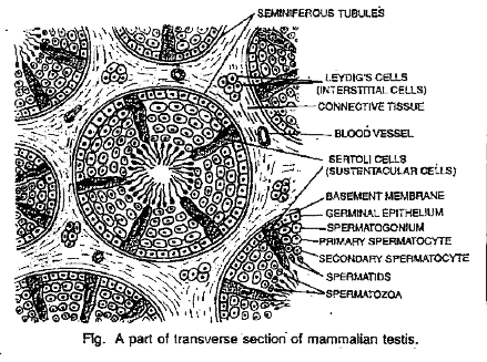
  3. Describe the structure of mammalian testis with the help of labelled d...

    Text Solution

    |

  4. What is Menstrual Cycle? Describe the role of FSH, LH, Oestrogens and ...

    Text Solution

    |

  5. Describe the ultra-structure of human sperm with the help of a labelle...

    Text Solution

    |

  6. What is spermatogenesis ? Briefly describe the process of spermatogene...

    Text Solution

    |

  7. What are foetal membranes. Give their names. Mention the function of ...

    Text Solution

    |

  8. Describe human female reproductive system with labelled diagram.

    Text Solution

    |

  9. Explain the role of the following hormones with reference to Menstrual...

    Text Solution

    |

  10. Describe the formation and fate of three germlayers inamammalian embry...

    Text Solution

    |

  11. At which stage of life does oogenesis begin in human females.

    Text Solution

    |

  12. Namethe organ where oogenesis gets completed inhuman females.

    Text Solution

    |

  13. Give a schematic representation of oogenesis in human females.

    Text Solution

    |

  14. What do you call the area of a human ovum from where the sperm makes t...

    Text Solution

    |

  15. Name the enzyme produced by the sperm to facilitate its entry.

    Text Solution

    |

  16. Draw a well labelled diagram of structure of Human ovum.

    Text Solution

    |

  17. Describe in detail the accessory glands of male and female humanbeing.

    Text Solution

    |

  18. What is cleavage ? Write down its characteristics.

    Text Solution

    |

  19. Differentiate between spermatogenesis and oogenesis.

    Text Solution

    |

  20. Explain the role of pituitary and the ovarian hormones in menstrual cy...

    Text Solution

    |