Text Solution
Verified by Experts


Topper's Solved these Questions
TISSUE AND TISSUE SYSTEM
PREMIERS PUBLISHERS|Exercise OTHER IMPORTANT QUESTIONS & ANSWERS (CHOOSE THE CORRECT ANSWER)|40 VideosTHE LIVING WORLD
PREMIERS PUBLISHERS|Exercise OTHER IMPORTANT QUESTIONS & ANSWERS (Answer the following questions.)|52 VideosTISSUE LEVEL OF ORGANISATION
PREMIERS PUBLISHERS|Exercise SOLUTION TO TEXTUAL QUESTIONS |6 Videos
Similar Questions
Explore conceptually related problems
PREMIERS PUBLISHERS-TISSUE AND TISSUE SYSTEM-OTHER IMPORTANT QUESTIONS & ANSWERS (ANSWER THE FOLLOWING)
- Write down the differences between tracheids and fibres.
Text Solution
|
- Give a brief answer on subsidiary cells in plant leaves.
Text Solution
|
- Explain the term trichomes.
Text Solution
|
- What do you understand about hypodermis in plant tissue system.
Text Solution
|
- What is pith?
Text Solution
|
- Explain the piliferous layer as epiblema.
Text Solution
|
- What is meant by stele in plant stem?
Text Solution
|
- Explain the nature of phloem in dicot stem.
Text Solution
|
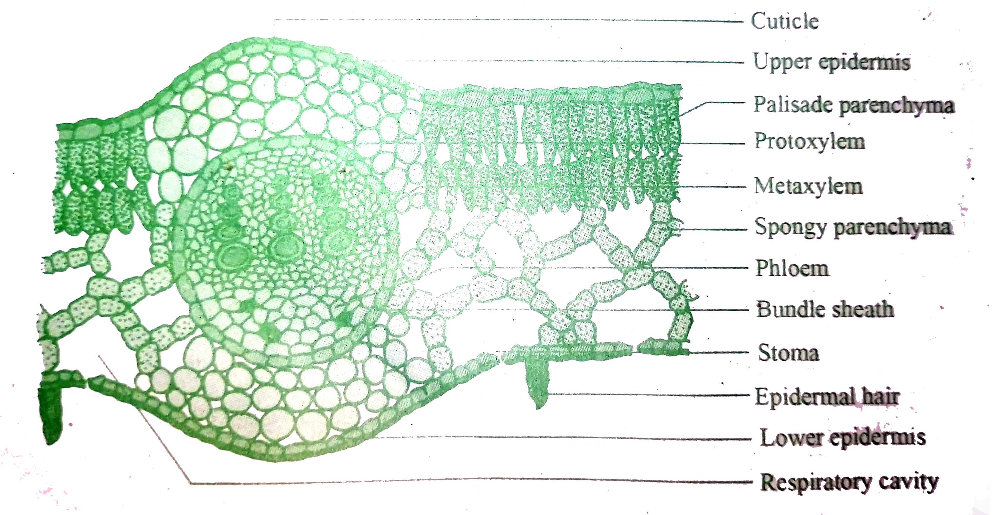
- Explain the mesophyll layer of leaf.
Text Solution
|
- Mention any three differences between stomata and hydathodes.
Text Solution
|
- What are halophiles?
Text Solution
|
- Answer in detail. Explain Histogen theory,
Text Solution
|
- Describe the structure and function of different kinds of parenchyma t...
Text Solution
|
- Describe the types of tracheids with diagram.
Text Solution
|
- What are the different types of plant tissues?
Text Solution
|
- Compare the vascular tissues of plant.
Text Solution
|
- Draw & label the ground plan of T.S of Dicot root
Text Solution
|
- Describe the vascular bundles of monocot stem.
Text Solution
|
- Draw and label the transverse section of monocot stem.
Text Solution
|
- Explain the parts of a flower, with a neat labelled diagram.
Text Solution
|