Transverse section of monocot or isobilateal leaf shows three parts - epidermis, mesophyll and vascular bundles.
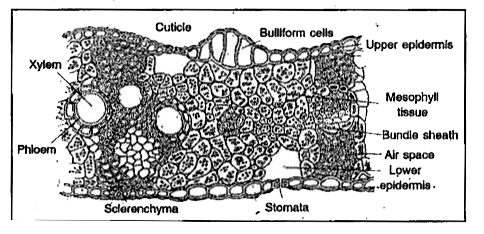
I. Epidermis:
1) This is the outermost layer on both sides of the leaf. . Cells are one cell in thickness. They are barrel shaped and closely packed without intercellular spaces.
2) It is covered by a waxy layer called cuticle.
3) Epidermis’on adaxial (upper) surface is called upper epidermis. Epidermis on abaxial (lower) surface is called lower epidermis.
4) Hairs are absent. Stomata are present on both sides in equal numbers.
5) in grasses specialised cells are present in upper epidermis. They are called bulliform cells or motor cells. They are thin walled and filled with water. They help in rolling and unrolling of the leaf.
6) Epidermis gives protection to inner tissues. Cuticle regulates transpiration. Stomata help in exchange of gases.
II. Mesophyll :
1) Ground tissue present between two epidermal layers is called mesophyll. It is chlorenchymatous.
2) Mesophyll is undifferentiated.
3) Cells have chloroplasts and perform assimilation of carbohydrates.
4) Sometimes patches of sclerenchyma are found beneath the epidermis. They provide mechanical strength.
III. Vascular Bundles :
1). Vascular bundles are present in the mesophyll in the form of veins.
2) Vascular bundles are conjoint, collateral and endarch.
3). Xylem is present on the upper side and phloem is present on the lower side.
4) Veins .help in conduction of water, mineral salts and food materials: They also provide mechanical strength.
5) Each vascular bundle is enclosed by a layer of special mesophyll cells called bundle sheath or border parenchyma.
6) Cells present on eitherside of vascular bundles towards upper and lower epidermis are called bundle sheath extensions. In many monocots, they are sclerenc hymatous and provide mechanical support.


